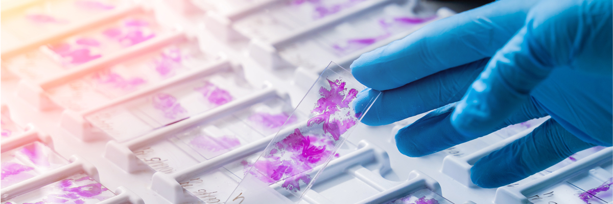
Aragen Bioscience’s Histology Services deliver precise tissue preparation and staining, crucial for detailed morphological analysis. Leveraging advanced techniques, we ensure exceptional quality and accuracy in your research and diagnostics. Trust our expertise to provide deeper insights into tissue structure and function, propelling your scientific and medical advancements forward.
Tissue processing
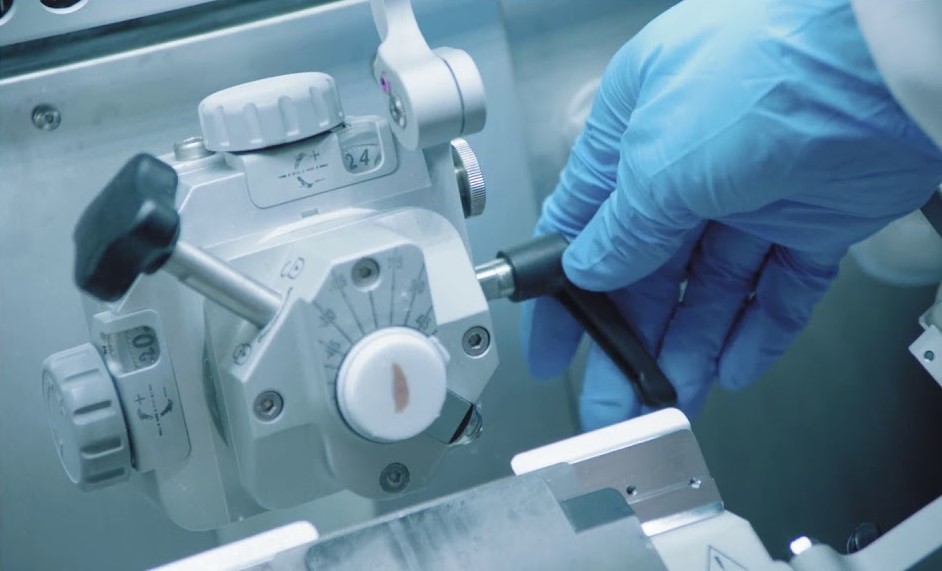
Tissue processing is the crucial first step in transforming biological samples into valuable insights, ensuring meticulous preparation that empowers our customers with precise and reliable results. Our tissue processing services encompass meticulous handling from trimming to sectioning, catering to both FFPE and fresh frozen samples.
Tissue microarrays construction
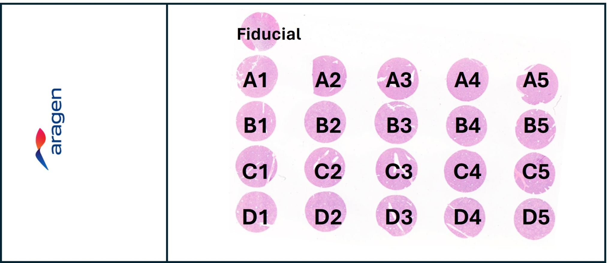
Tissue microarray (TMA) construction represents a pivotal advancement in research methodology, consolidating multiple tissue samples onto a single platform. This streamlined approach enables efficient analysis of biomarkers across diverse tissue types, fostering insights into disease mechanisms, treatment responses, and diagnostic markers with unparalleled efficiency and consistency.
At Aragen Bioscience, custom TMAs are expertly crafted to meet diverse research needs, drawing from our extensive library of samples including inflammation/fibrosis models, CNS disease models and CDX/Syngeneic tumor models.
Specialized and routine staining techniques
Specialized and routine staining techniques are pivotal in revealing intricate cellular details and identifying specific biomarkers, providing our customers with essential insights into tissue structure, function, and pathology. Enhance your tissue characterization with a comprehensive range of stains, from traditional H&E to specialized stains available with us.
General Tissues Stains
- Hematoxylin & Eosin (H&E): General tissue morphology, highlighting
nuclei and cytoplasm.
Connective Tissue Stains
- Masson’s Trichrome: Differentiates muscle, collagen fibers, and fibrin.
- Picrosirius Red: Collagen fibers
- Verhoeff’s Elastic Stain: Elastic fibers.
Neural Tissue Stains
- Luxol Fast Blue: Myelin in nervous tissue.
- Nissl (Cresyl Violet): Nissl bodies in neurons.
Hematological Stains
- Wright Giemsa: Blood smears, highlighting different blood cell types.
Pigment and Mineral Stains
- Fontana Masson: Melanin and argentaffin granules.
Carbohydrate Stains
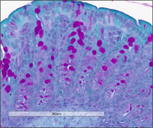
- Periodic Acid Schiff (PAS): Carbohydrates, glycogen, and mucin.
- Alcian Blue: Acidic polysaccharides like glycosaminoglycans.
Lipid Stains
- Oil Red O: Lipids in frozen tissue sections.
Tissue cross-reactivity assays (non-GLP)
Tissue Cross Reactivity (TCR) is a crucial preclinical assessment for therapeutic antibodies or antibody-like molecules required for regulatory submissions. It identifies potential off-target binding sites and evaluates the likelihood of unforeseen on-target interactions using frozen tissue samples. This informs decisions on animal model selection for assessing organ-specific toxicity in vivo, contributing essential data to the safety and specificity profiles of therapeutic candidates.
We design specialized assays to assess tissue specificity, suitable for research purposes that do not require GLP compliance.
- For therapeutic antibody or antibody-like molecule
- Safety assessment for early identification of off-target binding
- Species selection of animal toxicity models
Immunohistochemistry
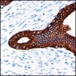
Immunohistochemistry (IHC) continues to evolve with advanced automated staining platforms, diverse antibodies across species, and novel biomarkers. These optimize staining protocols for precise target expression profiling in human and animal tissues, crucial for diverse studies like proliferation, immuno-oncology, and tumor profiling. Unlock the power of immunohistochemistry with our standard and customizable marker panels, supported by rigorous antibody screening and optimization.
RNAscope In-situ Hybridization
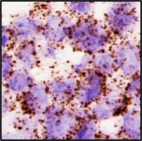
RNAscope In-situ Hybridization (ISH) revolutionizes molecular analysis by precisely localizing RNA molecules within tissue samples. This technique enables detailed spatial mapping of gene expression patterns, facilitating nuanced insights into cellular processes, disease mechanisms, and potential therapeutic targets with unparalleled specificity and sensitivity.
Explore with our experts the molecular landscape of tissues with RNAscope, offering single RNA molecule visualization in various sample types including FFPE and fresh frozen tissues.
Histopathology report
Pathology report writing support ensures clear and precise documentation of diagnostic findings, serving a diverse range of stakeholders beyond clinicians. This service facilitates effective communication of health information, supporting researchers, administrators, and patients alike in making informed decisions and improving overall health outcomes.
At Aragen Bioscience, insightful pathology reports generated by experienced board-certified pathologists ensure accurate interpretation of digital images.
